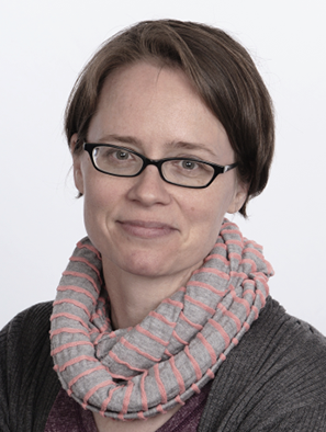
Dinah Loerke
Professor
What I do
Associate Professor with a focus on biophysics. We investigate the biomechanics of cells - particularly epithelial cell layers - using quantitative computational image and data analysis and modeling.Specialization(s)
Cell Biophysics, Biomechanics, High-Resolution Imaging, Computational Image Analysis
Professional Biography
Prof. Loerke completed her PhD degree in biophysics at the Max Planck Institute for Biophysical Chemistry in Gottingen (Germany) in 2004, and did her postdoctoral training in computer vision at The Scripps Research Institute (TSRI) in San Diego in the lab of Prof. Gaudenz Danuser, focusing on quantitative analysis of the temporal dynamics of clathrin-mediated endocytosis (in a collaboration with the lab of Prof. Sandra Schmid).
She joined the Department of Physics and Astronomy at DU in 2009. She was honored with an RCSA Cottrell Scholar Award (for early career teacher-scholars) in 2014, and was promoted to Associate Professor in 2016. Her current research interests revolve around the biomechanics of epithelial cell layers, particularly during morphogenetic movements in development, through the lens of computational analysis of dynamic live-cell imaging. Her work is collaborative and interdisciplinary, and has been supported by the National Institutes of Health (NIH), the Research Corporation for Science Advancement (RCSA), the Gordon and Betty Moore Foundation, and the National Science Foundation (NSF). Her teaching interests include biophysics, medical physics, optics and biomechanics, Universal Design for Learning (UDL), and graduate student mental health.
She joined the Department of Physics and Astronomy at DU in 2009. She was honored with an RCSA Cottrell Scholar Award (for early career teacher-scholars) in 2014, and was promoted to Associate Professor in 2016. Her current research interests revolve around the biomechanics of epithelial cell layers, particularly during morphogenetic movements in development, through the lens of computational analysis of dynamic live-cell imaging. Her work is collaborative and interdisciplinary, and has been supported by the National Institutes of Health (NIH), the Research Corporation for Science Advancement (RCSA), the Gordon and Betty Moore Foundation, and the National Science Foundation (NSF). Her teaching interests include biophysics, medical physics, optics and biomechanics, Universal Design for Learning (UDL), and graduate student mental health.
Degree(s)
- Ph.D., Biophysics, University of Goettingen, 2004
- MS, Physics, University of Goettingen, 1998
Licensure / Accreditations
- Adult CPR/AED
- IRBNet Board SL-1 Lab Certification
Professional Affiliations
- American Society for Cell Biology
- American Physical Society
Research
My overarching research interest is the field of cellular biomechanics: We are asking how we can combine experimental and computational imaging tools to elucidate how molecular mechanisms generate forces, and how these forces produce the macroscopically observable movement of tissues, cells, or organelles. Quantitative live-cell imaging and image analysis approaches are key elements of all of our current projects. Aside from some side projects related to membrane traffic and/or particle detection and tracking (an area which was the focus of my early career), my current primary interest is the cellular biomechanics of epithelial monolayers:
(1) Epithelial tissue dynamics during the early development of the fruit fly drosophila melanogaster, specifically the mechanics of cell intercalation during the elongation of the germband (which produces the elongation of the head-to-tail body axis of the fly embryo). Our work in this area is a collaboration with Prof. JT Blankenship, a fly geneticist in the DU Department of Biological Sciences.
(2) Epithelial architecture in the external vasculature of the sea squirt botryllus schlosseri, and mechanical changes during vascular aging. Our work in this area is a collaboration with Prof. Megan Valentine, Prof. Tony deTomaso (both University of California at Santa Barbara), and Prof. Adriana Dawes (Ohio State University).
Our work in these areas is inherently collaborative. In the last 25 years, the combination of new high-resolution microscopy techniques, together with advances in fluorescence biomarkers for live-cell imaging, has produced a wave of innovation (including multiple Nobel Prizes) and has enabled unprecedented insights into the molecular dynamics inside living cells. Computational image analysis techniques have been a critical resource in this field to process increasingly large datasets and extract meaningful mechanisms from multiplexed imaging data.
Areas of Research
image analysis
computer vision
biomechanics
particle detection
particle tracking
epithelial monolayers
germband extension
planar polarity
Key Projects
- The Biomechanics of Intercalation in Germ-Bank Elongation
- Deconstructing The Cell's Mechanical Circuits
- Engagement of cell ratcheting during epithelial morphogenesis
- Sliding vertex behaviors during epithelial morphogenesis and tissue elongation
- Volumetric analysis of epithelial morphogenesis with high spatiotemporal resolution -- Resubmission
Featured Publications
Vanderleest, T. E., Xie, Y., Smits, C., Blankenship, J. T., & Loerke, D. (2022). Interface extension is a continuum property suggesting a linkage between AP contractile and DV lengthening processes. Molecular Biology of the Cell, 33(14).
Rodriguez, D., Taketa, D., Madhu, R., Kassmer, S., Loerke, D., Valentine, M., & De Tomaso, A. (2021). Vascular Aging in the Invertebrate Chordate, Botryllus schlosseri. Frontiers in Molecular Biosciences, 8.
Presentations
Loerke, D. (2023). Cell Mechanics in Cell Intercalation During Germ Band Extension. Morphogenesis in Animals & Plants: Search for Principles. Santa Barbara: Kavli Institute for Theoretical Physics.
Loerke, D. (2023). Image Analysis and Tissue Biomechanics in Morphogenesis. CU Boulder Biomedical Engineering Seminar Series. CU Boulder Department of Biomedical Engineering .
Awards
- 2017 Scialog Fellow, Research Corporation for Science Advancement
- KITP fellow, Kavli Institute for Theoretical Physics (KITP)
- KITP fellow, Kavli Institute for Theoretical Physics (KITP)
- 2017/2018 NSM Excellence in Research, University of Denver
- DU Career Chamption Nominee, University of Denver